Digital Infrared Thermal Imaging “DITI” is a 15 minute non invasive test of physiology. It is a valuable procedure altering your doctor to change that can indicate early stage breast disease.
The benefit of DITI testing is that it offers the opportunity of earlier detection of breast disease than has been possible through breast self examination, doctor examination or mammography alone.
DITI detects the subtle physiologic changes that accompany breast pathology, whether it is cancer, fibrocystic disease, an infection or a vascular disease. Your doctor can then plan accordingly and lay out a careful program to further diagnose and/or MONITOR you during and after any treatment.
PROCEDURE
- Non invasive
- No radiation
- Painless
- No contact with the body
- F.D.A registered
This quick and easy test starts with your medical history being taken before you partially disrobe for the scanning to be performed. This first session provides the baseline of your “thermal signature”. A subsequent session assures that the patterns remain unchanged.
All of your thermograms (breast images) are kept on record and once your stable thermal pattern has been established any changes can be detected during your routine annual studies
WHO?
All women can benefit from DITI breast screening. However, it is especially appropriate for younger women (30 – 50) whose denser breast tissue makes it more difficult for mammography to be effective. Also for women of all ages who, for many reasons, are unable to undergo routine mammography. This test can provide a ‘clinical marker’ to the doctor or mammographer that a specific area of the breast needs particularly close examination.
It takes years for a tumor to grow thus the earliest possible indication of abnormality is needed to allow for the earliest possible treatment and intervention. DITI’s role in monitoring breast health is to help in early detection and monitoring of abnormal physiology.

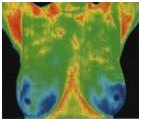
NORMAL
Good thermal symmetry with no suspicious thermal findings. These patterns represent a baseline that won’t alter over time and can only be changed by pathology.

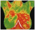
FIBROCYSTIC
Significant vascular activity in the left breast which was clinically correlated with fibrocystic changes.

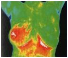
INFLAMMATORY CANCER
There were no visible signs of abnormality. Referral to a breast specialist and a subsequent biopsy diagnosed inflammatory breast cancer at a very early stage.

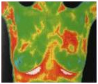
DUCTAL CARCINOMA
The vascular asymmetry in the upper left breast was particularly suspicious and clinical investigation indicated a palpable mass. A biopsy was performed and a DCIS of 2 cm was diagnosed.
 Alternative Primary Care


